
Atypical Nevus
Introduction
Atypical nevus is an unusual looking mole with irregular features when viewed under a microscope. It is commonly called Dysplastic Nevus or Atypical mole. These moles are not cancerous but need to be monitored closely. This is because individuals with these moles have a higher risk of developing Melanoma – a dangerous skin cancer, somewhere else on their body.
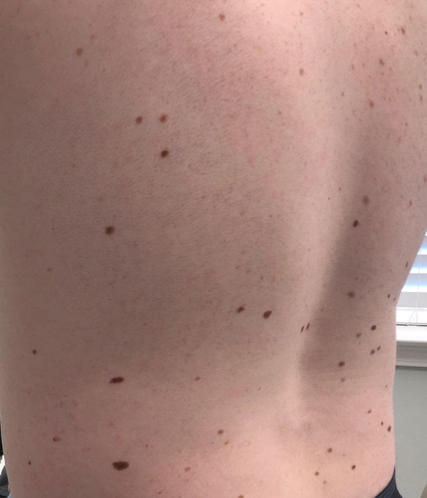
Atypical nevus can appear anywhere on the body, hence, it is very important to have thorough skin exams looking at every square inch of skin surface area. Look out for changes in the appearances of moles on your skin and call your dermatologist’s attention to any changes (growth, bleeding, asymmetry, itching, pain)
To identify an atypical nevus on your skin, look out for the following features when having self-examination: a size > 6mm in diameter, irregular borders, an unusual shape, different colors of the flat and bumpy parts, usually pink, brown and black. Unfortunately, these features overlap with melanoma and it important to have a professional at examine any new or concerning lesions to determine what they are.
EPIDEMIOLOGY
The United States population has about a 10% incidence of atypical nevi. However, a more accurate prevalence estimate is between 2 and 8 out of every 100 young, fair-skinned adults. According to the results of a study on dysplastic nevi carried out in families, patients with this condition have an elevated risk of melanoma. The risk gets even higher if there is a personal or family history of melanoma, or if numerous family members are affected. Increased sun exposure also results in increase in nevus development, although some nevi develop in sun-protected sites, like between the toes. Some genetic syndromes have predisposition to melanoma, breast cancer, pancreatic cancer, mesothelioma, and others, so its important to give your Pennsylvania Dermatology Specialist an accurate family history. The result shows that “the higher the number of dysplastic nevi, the greater the chances of having melanoma.”
"Look out for changes in the appearances of moles on your skin and call your dermatologist's attention to any changes (growth, bleeding, asymmetry, itching, pain)"

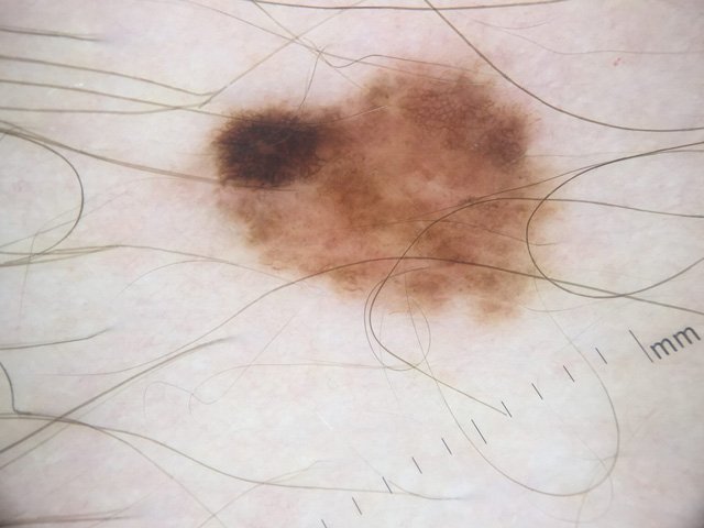
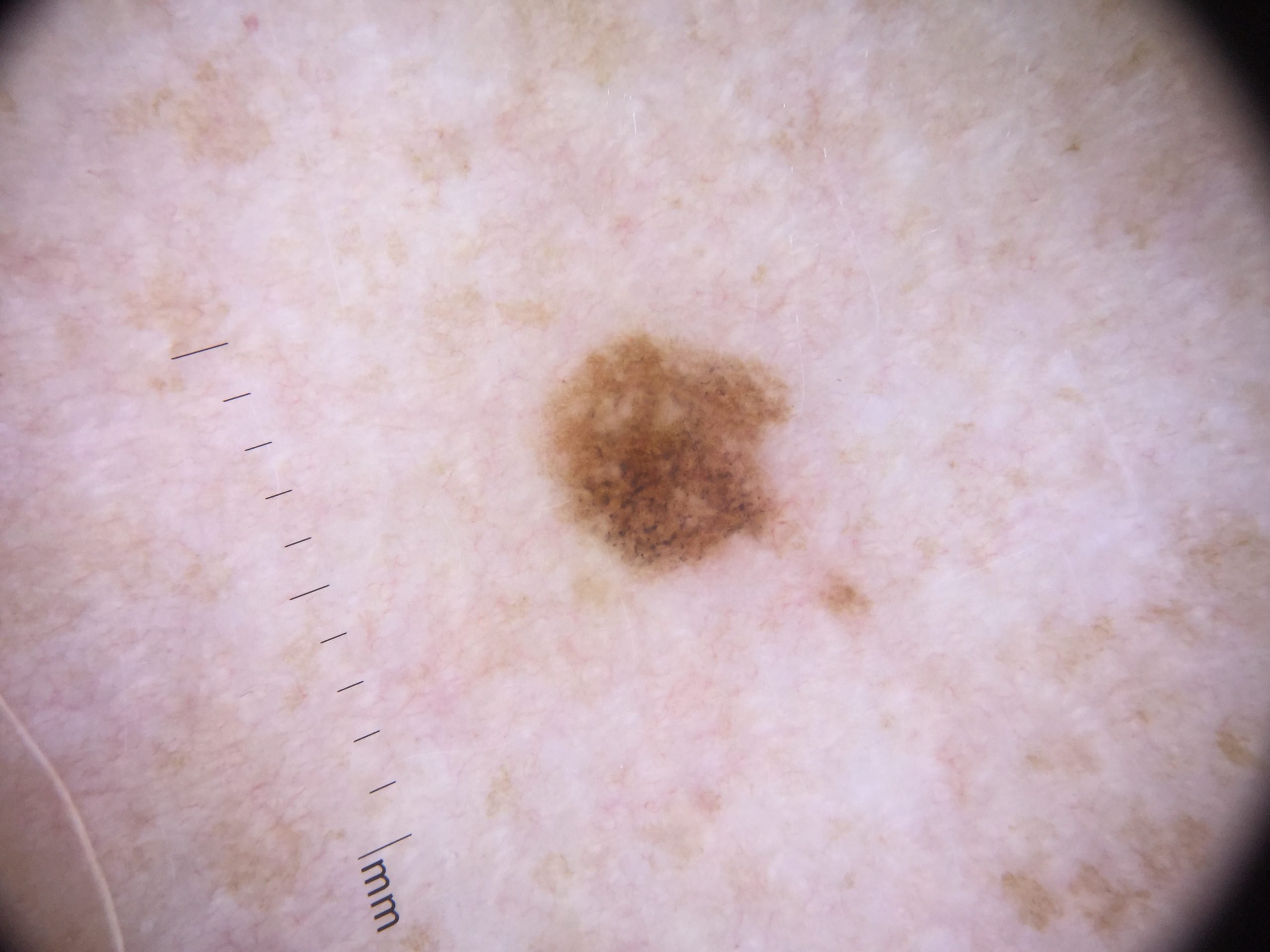
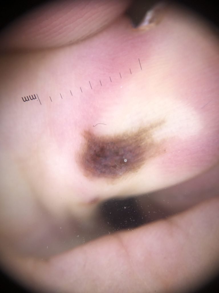
Dermoscopy is incredibly useful for evaluating Atypical Nevi. Both lesions above were dysplastic nevi.
SIGNS AND SYMPTOMS
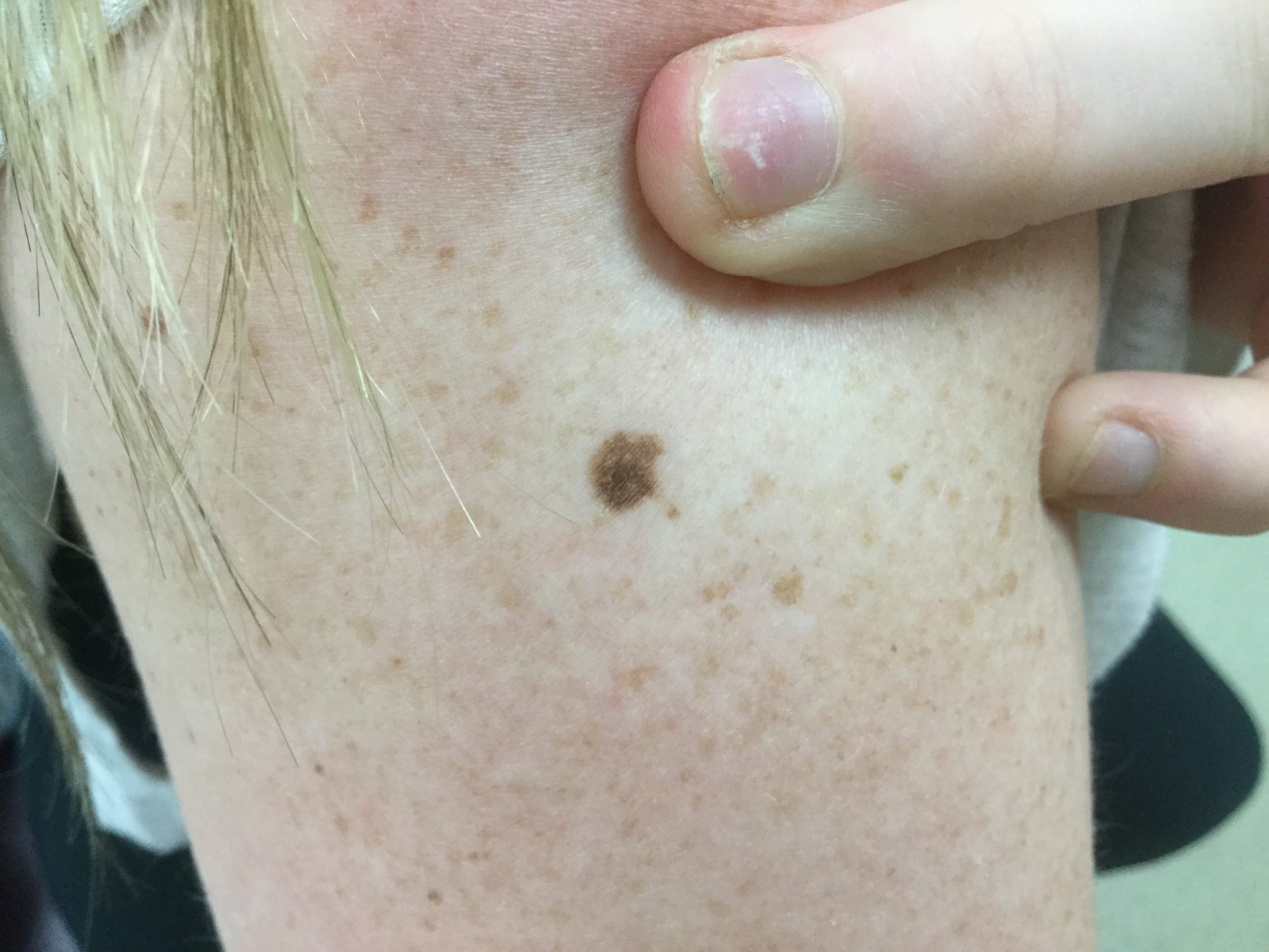
Atypical moles may appear anywhere on the skin. The lesions usually vary in size and/or color.
They can be greater than 6 mm and may vary in color ranging from pink to reddish-brown to dark brown.
Atypical moles may be darker brown in the center or periphery.
Proper viewing of the skin is done by using a piece of hand-held equipment called a dermatoscope. This can detect the high-risk features of cancer that have not yet been visible clinically. Using dermoscopy allows for the earliest diagnosis of a melanoma, before it has yet to develop the ability to spread (“caught in time”)
Patients with multiple areas of atypical moles and a personal or family history of Melanoma should be examined regularly as they are more prone to developing a Melanoma.
TREATMENT
Atypical nevus as earlier stated is not cancerous. However, it is advisable for patients to go for a complete skin examination. Patients should also be taught self-examination to identify any changes that may require immediate attention and treatment.
Surgical removal of the mole with a 2-3mm normal skin margin is often done for dysplastic nevi that have severe atypia under the microscope. The aim of the removal is to ensure that the lesion does not develop into melanoma. From studies, it has been shown that about 25-50% of melanomas arise from a pre-existing atypical nevus, so close monitoring is important.
Furthermore, surgical removal can lead to formation of scars which would be a concern especially if it’s in areas of cosmetic importance like the face. Therefore, a Pennsylvania Dermatology Specialist provider should determine the moles to be removed and the ones to be monitored in the most minimally invasive manner possible.
SkinIO skin imaging is a method of managing patients with numerous dysplastic nevi. It allows for 100% skin surface imaging with computer assisted algorithms allowing measurement of all of a patient’s nevi, follow-up in the future to determine change, and artificial intelligence for image assistance and dermoscopy. Ask your provider and schedule an appointment today!