
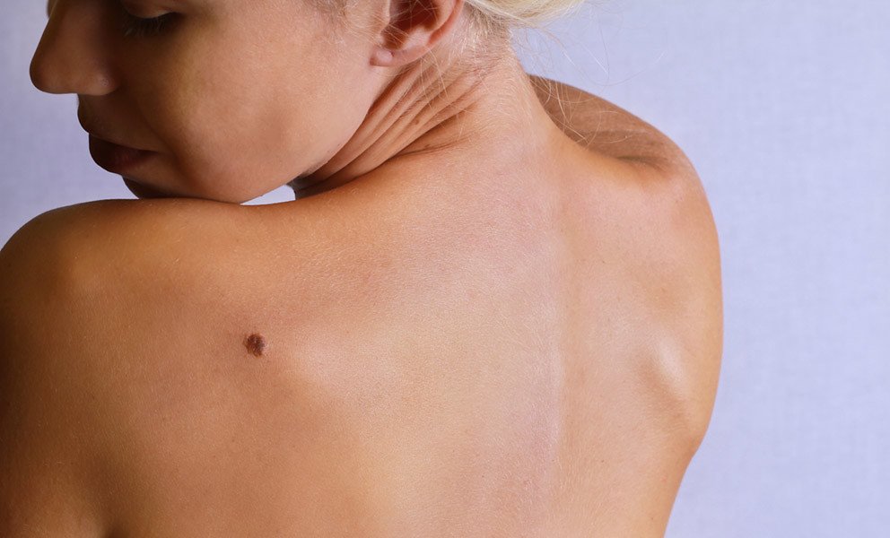
Biopsy of Skin Lesions
Wound care is fairly straightforward. Just cleanse the site(s) 1-2x/day with soap and water and cover with dressing and bandage. For dressing, we usually ask that you use Vaseline® or plain petroleum jelly. The reason is that we occasionally see patients who experience an allergic reaction to Neosporin® or bacitracin, but if you’ve used them in the past without problem, then it’s fine to use either instead of Vaseline®.
After you leave today, we will send the biopsy sample to the lab for evaluation. The results are typically available within several days. We will either discuss those results with you when you come back for follow-up or we will call you with the results if no follow-up has been scheduled.
Please keep in mind that a biopsy is simply a test. If the lab determines that the lesion contains abnormal or cancerous cells, additional treatment may be needed in order to ensure that no harmful cells remain.
Actinic keratoses (AKs)
Actinic keratoses (AKs) are often referred to as ‘pre-cancers’ since they carry an increased risk of turning into a squamous cell carcinoma. They don’t turn into melanoma. Overall, it’s a good thing for the lab to tell us that a lesion is ‘just’ an actinic keratosis since it means that the lesion hasn’t yet turned into a skin cancer. Treatment of these usually consists of freezing with liquid nitrogen or sometimes a topical cream can be prescribed.
Basal cell carcinoma (BCC)
Basal cell carcinoma (BCC) is sometimes referred to as the ‘best kind of skin cancer to have’ since they don’t tend to go too deep or ‘spread’ to other areas. Although they don’t spread, if left untreated they can eventually break down the skin and destroy the underlying and surrounding tissue. If caught in the early ‘superficial’ stage, they can be treated with a cream which acts like a topical form of chemotherapy. Most BCC, though, require excision or a procedure called electrodesiccation and curettage – a procedure in which the BCC is shaved off, remaining cells are ‘scraped away’, and the base is electrocauterized (burned).
Squamous cell carcinoma (SCC)
Squamous cell carcinoma (SCC) is the other ‘non-melanoma’ type of skin cancer. Although they have a potential to spread, it’s very rare for them to do so. The more common concern is that, similar to BCC, they can damage the underlying and surrounding tissue if left untreated. Treatment options are similar to those described for BCC.

Dysplastic or ‘atypical’ nevus
Dysplastic or ‘atypical’ nevus is the term given to a mole which is atypical but not yet melanoma. When the lab provides this diagnosis, they will classify the degree of atypia. A nevus which is classified as ‘mildly’ dysplastic/atypical is exhibiting minimal irregularity and poses very low risk of ever turning into a melanoma. Those which are ‘moderately’ or ‘severely’ dysplastic/atypical are more so and, although still not yet melanoma, carry a greater risk of eventually turning into skin cancer. When treatment is necessary, dysplastic/atypical nevi require either shave removal or excision.
Melanoma
Melanoma is the most well-known type of skin cancer. Because they represent cancer of the pigment cells (melanocytes), they typically present as a dark spot which keeps growing and changing. Although melanomas are actually the least common type of skin cancer, they’re potentially the most serious. If caught in the very earliest stages, they simply need to be excised in the office with no further treatment necessary other than regular re-evaluation and skin exams. If/when melanomas are diagnosed as being a little deeper, they do carry the risk of metastasizing – although most do not. Because of this risk, though, it may be necessary to not only excise, but also perform an evaluation of the lymph nodes just to be safe. Exactly which treatment approach is necessary for melanoma depends on many factors – including the depth and staging as determined by the pathologist.